Videos by Topic
To view videos in full-screen mode, click the full-screen arrows icon in the player. Videos will automatically play in full-screen mode on mobile devices.
Topics
AMD |
Instrumentation & devices |
Retinal detachments |
Diabetic retinopathy |
Intraocular lens |
Trauma |
Inflammatory disease |
Myopic foveoschisis |
AMD
 |
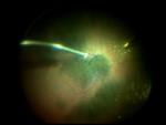
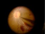
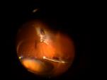
Subretinal TPAMauricio Maia, MD, PhD, and Rodrigo Milan Navarro, MD This video shows a patient with subfoveal hemorrhage due to polypoidal vasculopathy. He had already lost 1 eye due to past hemorrhage, and his best-eye visual acuity decreased to hand movements 1 day before surgery. The physicians decided to inject Tissue Plasminogen Activator (tPA) at a dose of 5 micrograms/0.1 cc using 0.2 cc of solution. |
 |
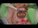
25-Gauge Vitrectomy for the Treatment of Massive Submacular HemorrhageShlomit Schaal, MD, PhD; Niloofar Piri, MD; Brooke Nesmith, MD
The case of an exudative-AMD patient with a vitreous hemorrhage and massive hemorrhagic retinal detachment. |
Diabetic retinopathy
 |
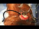
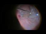
Vitreopapillary Traction: Managing the Posterior HyaloidHemanth Murthy, MBBS, MD, and Naveenam Srinivasa Muralidhar, MD Vitreopapillary traction is a rare complication of proliferative diabetic retinopathy. In this case, the posterior hyaloid is thickened and adherent to the retina posing difficulties in separation. Further, the optic nerve fibers are already affected by the traction.
|
 |
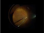
Diabetic Retinopathy: Current Techniques Using the Latest TechnologiesVirgilio Morales-Canton, MD
Innovative recommendations for diabetic retinopathy cases, including modifications on fluidics and cutting speeds and chandelier illumination using only a laser probe in order to perform adequate coagulation. |
Inflammatory disease
 |
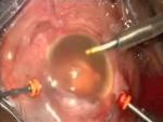
2-for-1: Removal of 2 Dislocated Fluocinolone Acetonide Implants With Placement of a New ImplantGlenn C. Yiu, MD, PhD
A complex procedure in which the surgeon uses the same sclerotomy to remove 2 fluocinolone implants in exchange for a new implant. |
Instrumentation & devices
 |
Macular Surgery: Techniques, Results and ComplicationsMarcelo Zas, MD, PhD, and Arturo A. Alezzandrini, MD, PhD This is a retrospective chart review of 32 consecutive 23g and 25g cases. The indications for surgical intervention were macular hole (stage 4) (n = 22) and macular epiretinal membrane (MEM) (n = 10). Brilliant Blue G and Tripan Blue were used to stain the membranes.
|
 |
23-Gauge PPV With ILM Peel for Fovea-Involving Sub-ILM Hemorrhage Due to Retinal Arterial MacroaneurysmEnchun M. Liu, MD, and Matthew A. Thomas, MD An 80-year-old woman presented with acute vision loss in her right eye due to a retinal arterial macroaneurysm. Because she had a large fovea-involving sub-internal limiting membrane (ILM) hemorrhage precluding a clear view of the subfoveal space, she elected for vitrectomy with ILM peel with the possibility of subretinal tissue plasminogen activator (tPA) injection if subfoveal hemorrhage was found. |
 |
Tough Hyaloid Forceps TechniqueJohn W. Kitchens, MD A demonstration of a difficult posterior hyaloid separation utilizing a forceps technique.
|
 |
Small-Gauge Retinal BiopsyJune 2014 Featured Video Baseer Ahmad, MD, and Bradley T. Smith, MD June 10, 2014
An example of 23-gauge vitreous and retinal biopsy taken from an eye with suspected primary vitreoretinal lymphoma. The use of air infusion for vitreous biopsy is illustrated, as well as the use of small-gauge diathermy, vertical scissors, and forceps for removal of the retinal tissue. |
 |
Lutein: A New Dye for ChromovitrectomyMauricio Maia, MD, PhD, and Rodrigo Milan Navarro, MD
This is a chromovitrectomy technique using a new green dye that improved the intraoperative identification of internal limiting membrane (ILM) in blue color, similar to Brilliant Blue G, and the posterior hyaloid/vitreous base identification in a golden appearance by deposition of lutein golden crystals. |
 |
ERM Removal Using a 27g Vitrectomy SystemJohn W. Kitchens, MD This is an assessment of a 27g system in a case of a patient with a previous posterior vitreous detachment and a taut epiretinal membrane. |
 |
Phaco Instrumentation for Pars Plana LensectomyApril 2014 Featured Video April 8, 2014
As demonstrated in this video, contemporary phacoemulsification instrumentation allows for more efficient and rapid removal of a luxated cataractous crystalline lens or lens fragments.
Several simple modifications allow for the implementation of this widely available anterior segment surgical instrumentation to pars plana lensectomy. |
 |
Epiretinal Membrane and Internal Limiting Membrane Peel Utilizing Wide-Field Viewing System With HD LensJohn W. Kitchens, MD February 11, 2014
This video demonstrates an epiretinal membrane (ERM) and internal limiting membrane (ILM) peel utilizing an HD lens. Despite the wide field of view provided by the lens, it provides great depth of field.
|
 |
Wide-Field Viewing System With HD Lens for Epiretinal Membrane Peel in a Patient With a Small PupilJohn W. Kitchens, MD February 11, 2014
Peeling an epiretinal membrane (ERM) in a patient with a small pupil is typically difficult, but viewing through an HD lens provides an optimal depth of field while also maintaining a wide field of view.
This video shows the utilization of an HD lens in the case of a male patient on Flomax (tamsulosin, Boehringer Ingelheim Pharmaceuticals, Inc, Downers Grove, IL) with a horseshoe-shaped tear as a result of an ERM.
|
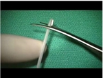 |
New Intraocular Foreign Body ForcepsNur Acar, MD, FEBO View newly designed intraocular foreign body (IOFB) prototype forceps and watch simulations of their use in IOFB removals.
|
 |
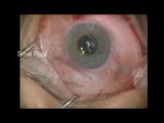
Illuminated External Needle DrainageJohn W. Kitchens, MD
A new technique for subretinal fluid drainage using both a guarded needle and a 29-gauge chandelier.
|
Intraocular Lens
 |
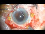
Intraocular Lens Refixation Using GORE-TEX SutureJuly 2014 Featured Video This is a case of a refixation of a partially dislocated CZ70BD lens that had been sutured to the sclera with Prolene (polypropylene sutures, Ethicon Endo-Surgery, Inc, Somerville, NJ) 10 years ago. One of the Prolene sutures had broken, while the other was still intact.
|
 |
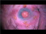
Scleral Fixated Intraocular Lens Using GORE-TEX Suture and Akreos LensApril 2014 Featured Video Omesh P. Gupta, MD, MBA; Marc J. Spirn, MD; and Jason Hsu, MD April 8, 2014
This is a technique of scleral fixating a Bausch + Lomb Akreos Lens using GORE-TEX suture. The attractive features of this approach are the use of standard small incision vitrectomy instrumentation and a standard phacoemulsification wound.
|
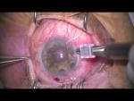 |
Novel Technique for IOL RescueFebruary 2014 Featured Video Jonathan L. Prenner, MD; Harold M. Wheatley, MD; and John D Wilgucki, BS February 11, 2014
An eye in which a secondary intraocular lens (IOL) completely dislocates into the posterior segment while remaining completely in the bag presents a unique surgical challenge. One can remove the lens/bag complex and place a secondary IOL or explant the lens from the bag and then reposition the lens. The surgeons present a novel, minimally invasive procedure that allows for suture fixation of the lens/bag complex without the need for manipulating the conjunctiva.
|
 |
Bimanual IOL RescueOctober 2013 Featured Video This unique procedure allows for easier externalization of haptics and eliminates any anterior-segment manipulation. |
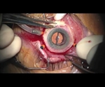 |
Sutureless Scleral Fixation of an IOLAugust 2013 Featured Video An inventive sutureless technique for secondary intraocular lens (IOL) rescue. |
Myopic foveoschisis
 |
Management of Myopic Foveoschisis With Vitrectomy With ILM PeelingHector H. Hernandez-Torres, MD, and Francisco J. Rodriguez, MD
An interesting approach incorporating Brilliant Blue G staining yields technical feasibility and good visual and anatomical outcomes. |
Retinal detachments
 |
Iatrogenic Retinal Detachment Inflicted During 25-Gauge Air-to-Fluid ExchangeOctober 2014 Video of the Month Christopher G. Fuller, MD This is the case of a 67-year-old woman who underwent vitrectomy for chronic vitreomacular traction and macular puckering. She suffered a retinal detachment at the time of surgical repair, likely secondary to intraocular irrigation reinitiated after Brilliant Blue G staining under air. It was the surgeon's first experience with Brilliant Blue G and provided an unexpected (and somewhat unhappy) teaching moment about the perils of air-to-fluid exchange.
|
 |
Retinotomies and Retinectomies: When and How? Current TechniquesSeptember 2014 Video of the Month Marcelo Zas, MD, PhD, and Arturo A. Alezzandrini, MD, PhD Retinotomies and retinectomies can improve the therapeutic effects of complicated retinal detachments (RDs) in proliferative vitreoretinopathy (PVR) cases. Complications of these maneuvers are high and should be managed properly.
|
 |
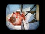
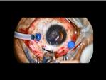
An approach to a Patient With Failed Cornea and Retinal Pathology -- Temporary Keratoprostheses and Pseudo-IrisYannek I. Leiderman, MD, PhD
This case illustrates the approach to a patient with a failed cornea and history of recurrent retinal detachment. Combined pars plana vitrectomy and penetrating keratoplasty were performed. A pseudo-iris was fashioned from a cyclitic membrane, conferring improved cosmesis and retention of silicone oil.
|
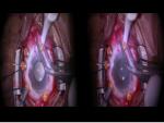 |
Stereoscopic 3D Video Demonstrating the Repair of a Complex Retinal DetachmentShlomit Schaal, MD, PhD; Augustina Palacio, MD; Douglas Sigford, MD; and Ahmet Ozkok, MD
This video illustrates the complex repair of a chronic rhegmatogenous retinal detachment with proliferative vitreoretinopathy. It was recorded using 2 different cameras, allowing 3-dimensional viewing. Video page includes 3D viewing instructions. |
 |
Diabetic Traction and Rhegmatogenous Retinal Detachment 27-Gauge VitrectomyMay 2014 Featured Video May 13, 2014
|
 |
Subretinal Band ExcisionMay 2014 Featured Video May 13, 2014
A case of a 40-year-old woman with a rhegmatogenous retinal detachment (RRD) proliferative vitreoretinopathy (PVR) and subretinal band along the inner aspect of the macula. This video explores and illustrates the decision-making process and intraoperative technique of the subretinal band excision.
|
 |
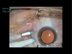
Traction and Rhegmatogenous Retinal Detachment: 25g Probe-Peel TechniqueMaria H. Berrocal, MD
|
 |
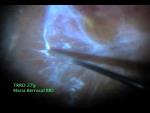
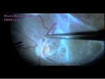
Diabetic Traction Retinal Detachment Repair Utilizing 7500 CPM 25-Gauge VitrectomyMaria H. Berrocal, MD
A demonstration of 25-gauge vitrectomy utilizing a 7500 cuts per minute (CPM) probe to remove fibrovascular tissue from the surface of the retina.
Using this method, the surgeon can safely perform peeling of the membranes with minimal traction on the underlying retina. |
 |
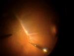
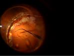
25-Gauge Pars Plana Vitrectomy of Total Retinal Detachment and Massive Fibrovascular ProliferationMaria H. Berrocal, MD
This video shows a case of fulminant progression of fibrovascular tissue in a young diabetic patient, managed with 25-gauge membrane peeling techniques and pre-operative anti-VEGF injection.
|
 |
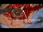
23-Gauge Retinectomy Under Silicone OilMarch 2014 Featured Video
This is an illustration of a method of performing retinectomy |
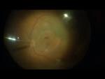 |
Management of a Macular Hole Secondary to a Retinal MacroaneurysmJohn W. Kitchens, MD
A case of a large macular hole secondary to a retinal arterial macroaneurysm that bled into the subretinal space and resulted in a full-thickness macular hole. |
 |
Tractional Retinal Detachment: 101Kevin J. Blinder, MD, and Enchun M. Liu, MD
This video shows a basic tractional retinal detachment/pre-macular hemorrhage—with a surprise at the end.
|
 |
Scleral Buckling IlluminatedJanuary 2014 Featured Video
Scleral buckling is an important technique for retinal detachment repair, and various approaches have been described. This video demonstrates the use of an operating microscope for superior visualization and better surgeon ergonomics. Using the BIOM (Insight Instruments, Inc, Stuart, FL) with an endoilluminator chandelier allows for intraoperative fundus visualization and assists with cryotherapy of retinal breaks and drainage of subretinal fluid. |
 |
Internal Limiting Membrane Peeling for Primary Rhegmatogenous Retinal DetachmentGaurav K. Shah, MD, and Rajesh C. Rao, MD
An innovative internal limiting membrane (ILM) peeling technique and highlights of a recent study that links ILM removal during primary retinal detachment repair to decreased secondary macular pucker formation. |
 |
Repair of Retinoschisis-Associated Retinal DetachmentSeptember 2013 Featured Video Gaurav K. Shah, MD, and Baseer Ahmad, MD
A demonstration of schisis, inner holes, and ILM peeling over a detached retina. |
 |
Retinectomy Under OilNovember 2013 Featured Video
View the difficult case of a 20-gauge retinectomy in an oil-filled eye of a patient with a proliferative vitreoretinopathy (PVR)-related redetachment. |
 |
Straight to OilJohn W. Kitchens, MD
In this case, a male patient presented with a retina re-detachment after silicone oil removal. Following removal of the dense cataract, the physicians decided to go directly to oil, rather than the more traditional approach of performing a retinectomy, utilizing perfluoro-n-octane (PFO) and then placing oil. |
 |
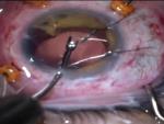
Abnormal Vitreous BaseGaurav K. Shah, MD, and Baseer Ahmad, MD This video shows an eye with an abnormal vitreoretinal interface with numerous iatrogenic intraoperative retinal breaks along the vitreous base. Laser cerclage is performed, and the surgeons demonstrate the utility of 360° laser and scleral buckling after vitrectomy. |
 |
Attacking PVRJohn W. Kitchens, MD
A variety of approaches are used to successfully reattach a retina, including anterior vitrectomy, membrane peeling, and peeling under perfluoro-n-octane (PFO). |
Trauma
 |
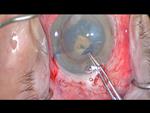
Evisceration or Reconstruction in a Case of Globe Rupture With No Light PerceptionS. Natarajan, MD; Juhi Garg, MBBS, MS; Divya Gupta Thakur, MBBS, MS; and Purva Valvekar, MBBS, MS A 40-year-old male presented with an injury from a bamboo stick. There was no light perception in the right eye, the patient had total hyphema with a flat anterior chamber, and CT showed a distorted globe. A B-scan showed posterior dislocation of the lens with dense vitreous hemorrhage. A large superior scleral tear extending up to the optic disc posteriorly with incarcerated vitreous was also noted.
|
 |
Pars Plana Retrieval of Glass Intraocular Foreign BodyRithwick Rajagopal, MD, PhD; Daniel P. Joseph, MD, PhD; and Rajendra S. Apte, MD, PhD
A man involved in a motor vehicle collision presented with a large retained intravitreal foreign body in the left eye, presumed to be glass based on the history of windshield shatter. Removal presents a challenge because of the large, irregular shape of the object and its nonmagnetic nature. |
 |
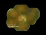
Four-Headed Retinal Surgical Monster: RP + ERM + FTMH + RDChristopher G. Fuller, MD
This is the case of a 65-year-old white woman with a long history of retinitis pigmentosa (RP) and best corrected visual acuity (BCVA) 20/60 in both eyes. After suffering a fall, she reported sudden vision loss in her left eye and presented with RP, full-thickness macular hole (FTMH), somewhat chronic-appearing epiretinal membrane, and macular-involving retinal detachment.
|
 |
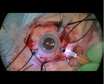
Microincisional Vitrectomy Instrumentation in Anterior and Posterior Segment Eye TraumaYannek I. Leiderman, MD, PhD This alternative to traditional anterior segment techniques utilizes vitrectomy cutting, aspiration, blunt dissection, and scissors capabilities of a vitrectomy probe, with minimal instrument exchanges. |
The Innovative Retina Surgical Video Series is made possible by a contribution from Alcon Laboratories, Inc.
Submit your teachable moments on video
Click here for full program details and video upload instructions.
|
The Innovative Retina Surgical Video Series |