

Retinoschisis
Degenerative retinoschisis (pronounced ret in oh SKEE sis) is an age-related condition in which the layers of the retina, the light-sensitive tissue at the back of the eye, begin to separate or split. Degenerative retinoschisis usually has no symptoms, can affect both eyes, and is detected by examination of the retina (Figure 1).
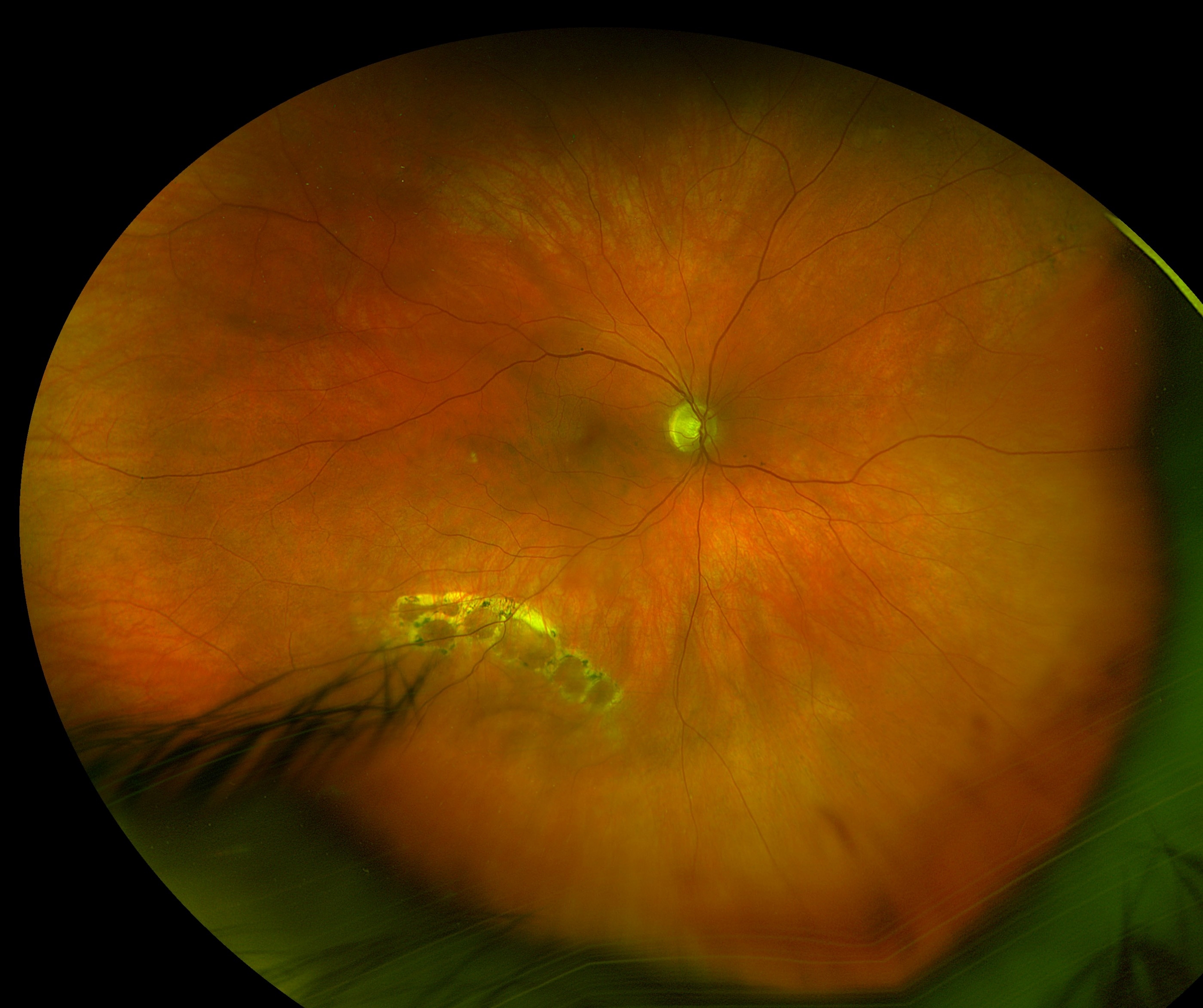
Figure 1. Retinoschisis with outer layer holes: The retinal separation can be difficult to see in this photograph. The lighter areas surrounding the small, long-standing holes help show the edge of the affected retina. |
 |
Symptoms
Degenerative retinoschisis often does not cause noticeable symptoms and is usually discovered during a routine dilated eye exam.
Your retina specialist may notice these signs:
- A smooth, clear area where the inner layers of the retina are raised or separated, without the wrinkles or folds typically seen with a retinal detachment.
- Lack of movement in the raised area when the eye moves.
- No full-thickness retinal tear, pigment cells or other signs associated with a retinal detachment.
Causes
As we age, tiny fluid-filled spaces can develop within the retina. This age-related change, called microcystoid degeneration, can cause some of the layers of the retina to separate or split.
Reticular retinoschisis is a less-common form of degenerative retinoschisis in which the separation occurs in the nerve layer of the retina and can be more pronounced. In some cases, reticular retinoschisis can progress to a full-thickness retinal tear, which may lead to a retinal detachment.
Studies suggest that degenerative retinoschisis occurs in approximately 4% of the population over the age of 40.
Diagnostic testing
Your retina specialist may use several tests to distinguish retinoschisis from a retinal detachment and to better understand the condition:
- Optical coherence tomography (OCT): This noninvasive imaging test creates detailed pictures of the retina. In retinoschisis, an OCT scan can show where the layers of the retina have separated, without fluid collecting underneath the retina (Figure 2).
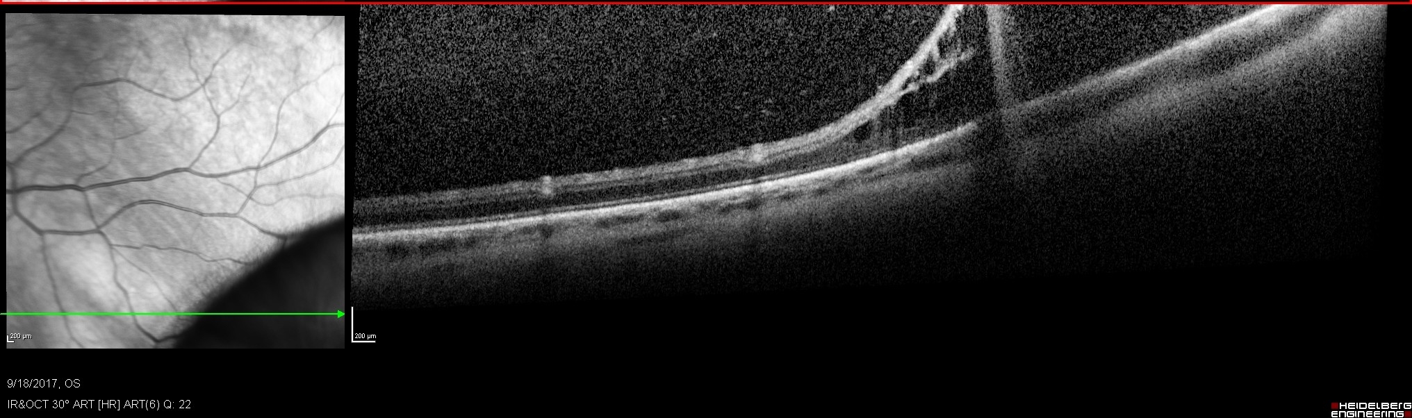
Figure 2. OCT of retinoschisis showing a splitting of the retina in the area of retinoschisis to the right. The green line to the left shows the position of the scan on the retina and this infrared image shows an area of black corresponding to the retinoschisis. |
- Laser testing: In some cases, a retina specialist may use a laser to help distinguish retinoschisis from a retinal detachment. Laser treatment normally produces a visible response in an area of retinoschisis. If there is fluid underneath the retina, as occurs with a retinal detachment, the laser may not produce the expected response.
- Visual field testing: This test measures areas of vision that may be reduced or missing. Retinoschisis typically causes a complete area of missing vision (an absolute scotoma), while a retinal detachment may cause a partial reduction in vision (a relative scotoma).
Treatment and prognosis
- Retinoschisis rarely causes loss of vision and should be closely monitored by your retina specialist with observation and retinal examination since the majority remain stable.
- If the retinoschisis progresses to a full-thickness rhegmatogenous retinal detachment, surgery is needed. Surgical options include pars plana vitrectomy with or without scleral buckle, or scleral buckle alone. Sometimes laser is placed as a barrier to try to prevent the retinoschisis from progressing but this treatment is not always successful. The surgical approach should be tailored to the ocular anatomy and location of the detached retina.