Hydroxychloroquine-Induced Retinal Toxicity
Hydroxychloroquine (HCQ), sold under the brand name Plaquenil, is a medication used to treat autoimmune diseases like rheumatoid arthritis and lupus. While it is very effective, long-term use can cause serious eye problems, including retinal damage and vision loss. About 7.5% of people on HCQ develop retinal issues, increasing to 20% after 20 years.
Symptoms
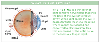
HCQ toxicity usually does not cause symptoms in the early stages. Patients typically do not notice any issues until the toxicity affects the central macula, a small area at the center of the retina where light is sharply focused to produce the detailed color vision needed for tasks such as reading and driving. When symptoms do appear, they include painless, progressive blurring of central vision.
Risk factors
Certain factors increase the risk of eye damage from HCQ:
- Daily dose over 6.5 mg/kg
- Obesity
- Use longer than 5 years
- Kidney or liver problems
- Age over 60
- Existing retinal disease
Screening: Eye damage from HCQ can occur before symptoms appear (Figure 1). Early and regular eye exams are crucial. Guidelines suggest a baseline exam within the first few months of starting HCQ. If no other risks are present, follow-up exams are recommended yearly after the first 5 years.
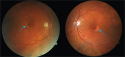
Figure 1 Fundus photos of the right and left eyes show pigment loss around the center of the retina. Photo courtesy of Lejla Vajzovic, MD, FASRS |
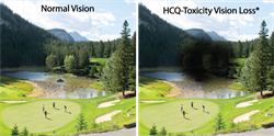
Figure 2 Simulated vision loss from HCQ retinal toxicity. *Vision loss looks different for each individual. Photo courtesy of Lejla Vajzovic, MD, FASRS |
Diagnostic testing
- Optical coherence tomography (OCT): Creates detailed images of the retina to detect early damage (Figure 3)
- Visual field testing: Measures how well different parts of the retina respond to visual stimuli
- Multifocal electroretinography (mfERG): Assesses retinal function in different areas of the retina
- Fundus autofluorescence (FAF): Non-invasive test that shows changes in retinal cells (Figure 4)
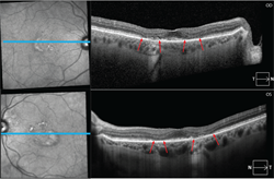
Figure 3 OCT images of the right and left eyes show damage to the outer retina indicated by disruption with loss of some of the outer layers of the retina in the bottom of the cross section in a patient with HCQ-induced maculopathy. Photo courtesy of Lejla Vajzovic, MD, FASRS |
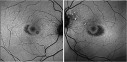
Figure 4 Fundus autofluorescence (FAF) images show dark areas around the center, creating what is described as a bulls-eye maculopathy since it looks like the center of an archery target. Photo courtesy of Lejla Vajzovic, MD, FASRS |
Treatment and prognosis
There is no treatment for HCQ-induced retinal damage. The best approach is early detection and stopping the medication if damage is found. Keeping the daily dose below 5.0 mg/kg can reduce the risk.
If eye damage is detected, switching to a different medication is recommended. After stopping HCQ, mild damage may improve. However, it is also possible that worsening could occur in the year following cessation, potentially leading to central vision loss.